


Case Study
Superoxide dismutase (SOD1) and the A4V mutation found in aggressive familial ALS.
Background
The amyotrophic lateral sclerosis disease (ALS) is a disorder characterized by the gradual degeneration and death of motor neurons. Approximately 10% of all ALS occurrences are genetically related and are inherited in an autosomal recessive manner. Specifically for such cases, the disease is named familial ALS (FALS). About 20% of FALS are directly linked to mutations found in superoxide dismutase (SOD1), a dimeric structure whose optimal functioning depends ultimately on how well two monomers are bound. The substitution of the alanine (at position) 4 by valine is a frequently found SOD1 mutation in aggressive forms of FALS.
Molecular dynamics studies have shown that this particular mutation produces less stable dimeric structures and destabilizes the metal-binding site, eventually leading to misfolded enzyme state. Since it was already known that the SOD1 uses electrostatic attraction to achieve faster than diffusion limited substrate approach and recognition, exploring even further the electrostatic component for both stability and substrate-related issues is important for understanding FALS. Here we present how MSSP can be used to help in such task.
Selecting the structures
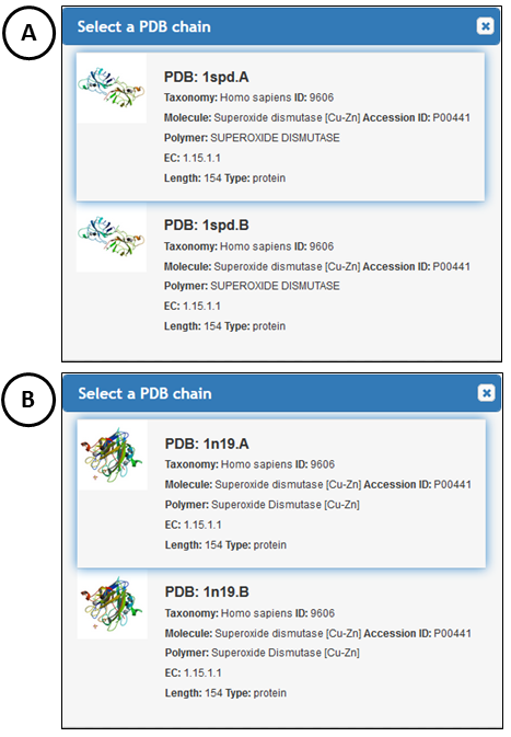
Select the PDB structure 1SPD and its chain A (wild type SOD1; Figure 1A) and 1N19, chain A (the SOD mutated structure: A4V but also containing substitutions of its two free cysteine residues: C6A and C111S; Figure 1B).
Reading the electrostatic potential
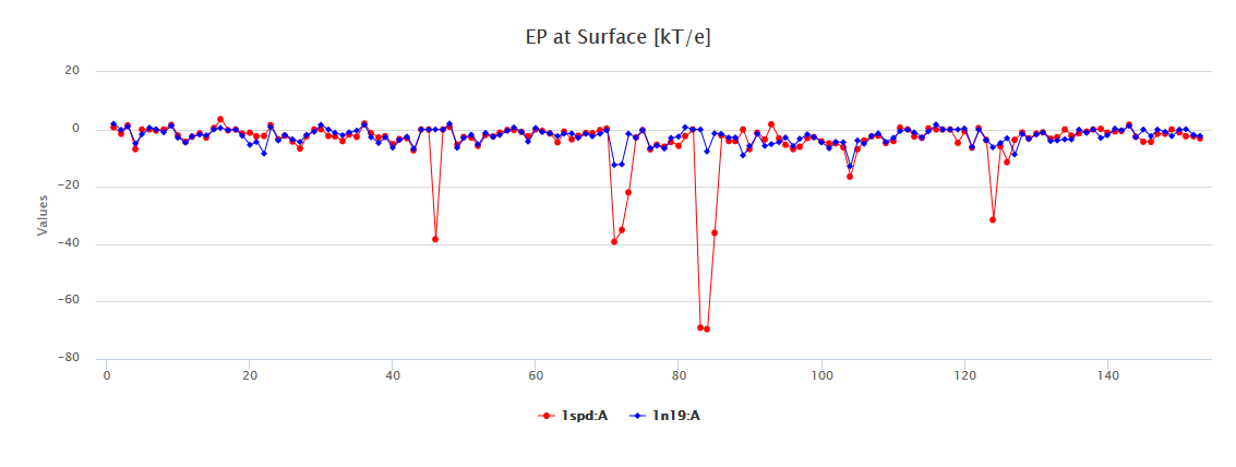
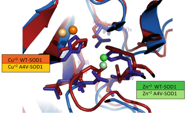
Launch one of the MSSP versions and then select the parameter Electrostatic Potencial @ Surface (EP@Surf). In Figure 2, one can observe that the mutated structure has a dramatic decrease of EP@surf at different positions, nevertheless, remote to the site of the mutated alanine (position number 4). A more thorough inspection of amino acid residues that suffered a great modification in value for their respective EP@surf reveals that they are involved and/or very close to the metal-binding atoms (Figure 3).
Back to Launching STINGAllo, or go to the Server.